The Science of Electrical Stimulation for Muscle Recovery: How It Works
Electrical stimulation for muscle recovery aids tissue repair by enhancing local microcirculation, accelerating metabolic waste removal, and directly engaging high-threshold fast-twitch muscle fibers to maintain contractile strength. By sending targeted electrical impulses through surface electrodes, this modality bypasses central nervous system fatigue to stimulate blood flow and preserve muscle function.
When we move naturally, our central nervous system sends electrochemical signals down the spinal cord to somatic motor neurons. These action potentials cross the neuromuscular junction, triggering the release of calcium within muscle cells and prompting actin and myosin filaments to slide together, creating a voluntary contraction.
Under natural voluntary recruitment (governed by Henneman’s size principle), the body recruits smaller, fatigue-resistant Type I (slow-twitch) motor units first. Only when load, speed, or fatigue demands it does the brain recruit larger Type II (fast-twitch) motor units.
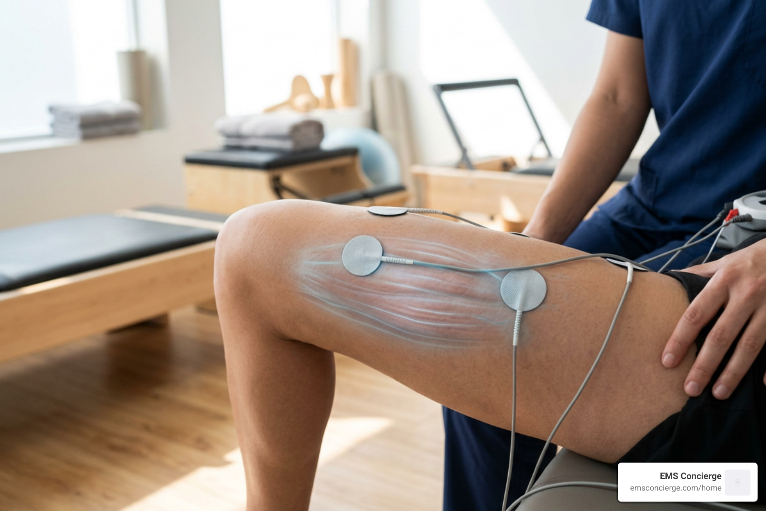
Electrical muscle stimulation turns this recruitment order on its head. When electrical impulses pass through surface electrodes into the underlying motor nerve branches, they depolarize axonal membranes directly (Blazevich et al., 2021). Because large-diameter fast-twitch motor axons have lower internal electrical resistance, electrical stimulation preferentially or synchronously engages Type II motor units alongside Type I units, even at submaximal force outputs.
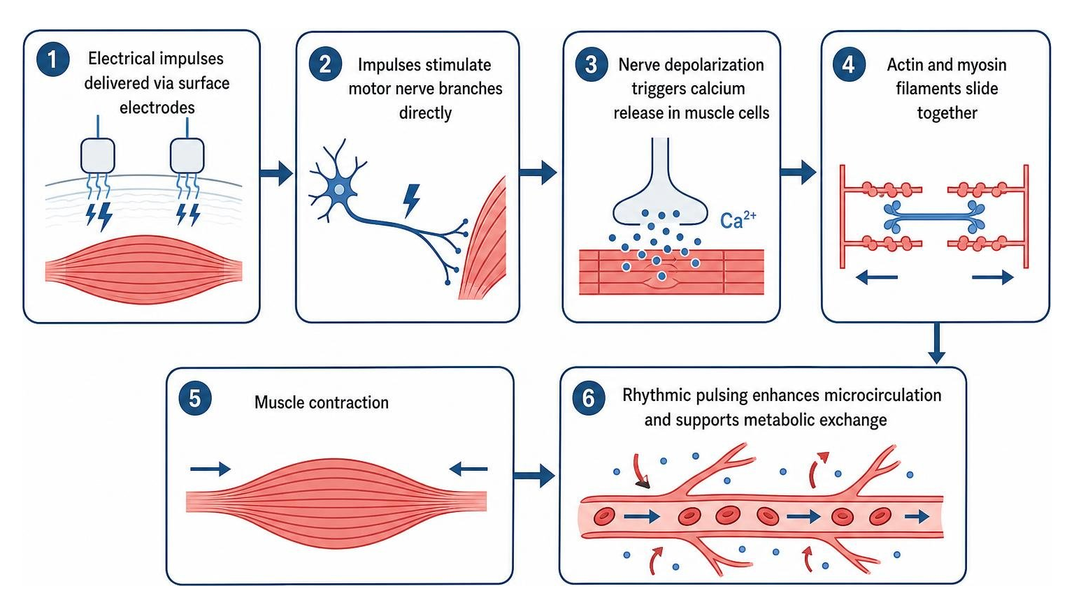
This non-selective, synchronous recruitment activates muscle fibers that often remain dormant during low-intensity voluntary movements. Furthermore, rhythmic electrical pulsing acts as a mechanical pump on the surrounding microvasculature, increasing local blood flow and promoting metabolic exchange in targeted tissues.
Modality Differences: NMES, TENS, and Microcurrent (MENS)
Not all electrical currents serve the same purpose. Understanding how different waveforms interact with human physiology helps clarify what each tool can achieve:
- Neuromuscular Electrical Stimulation (NMES / EMS): Operates at higher intensities designed to cross the motor threshold, directly depolarizing motor axons to evoke visible, rhythmic muscle contractions. It is primarily used to counteract disuse atrophy, improve motor unit recruitment, and enhance muscle activation.
- Transcutaneous Electrical Nerve Stimulation (TENS): Focuses on sensory nerve fibers rather than motor nerves. By sending low-intensity, non-contractile signals to cutaneous sensory receptors, TENS activates the "gate control" mechanism in the spinal cord, dampening nociceptive pain signals traveling to the brain (Keriven et al., 2025). A systematic review of 381 randomized trials involving 24,532 participants demonstrated that pain intensity was significantly lower during and after TENS treatment compared with placebo.
- Microcurrent Electrical Neuromuscular Stimulation (MENS): Delivers sub-sensory currents (measured in microamperes, $mutext{A}$) that mimic the body’s endogenous bioelectric fields. Research into acute microcurrent electrical stimulation on muscle function reveals that applying low-intensity microcurrent can alter oxygen kinetics, accelerating deoxyhemoglobin clearance in local microvasculature while shifting the autonomic nervous system toward parasympathetic recovery (Piras et al., 2021).
| Modality | Target Tissue | Primary Output | Physiological Objective |
|---|---|---|---|
| NMES / EMS | Motor Axons | Visible Muscle Contraction | Strength preservation, neuromuscular re-education, hyper-recruitment |
| TENS | Sensory Nerve Fibers | Tingling Sensation (No Contraction) | Gate-control pain modulation, analgesia |
| MENS | Cellular / Microvascular | Sub-Sensory (No Sensation) | Microvascular perfusion, ATP synthesis, autonomic recovery balance |
Cellular and Hemodynamic Mechanisms
At the cellular level, the mechanical tension evoked by electrical pulses triggers downstream intracellular signaling pathways, such as the mTOR pathway, which regulates muscle protein synthesis.
During standard disuse, fast-twitch (Type II) fibers atrophy rapidly. Because electrical stimulation targets these higher-threshold fibers directly, it helps maintain contractile protein density within muscle fibers during periods of restricted mobility (Cheuy et al., 2023). Research into enhancing adaptations to NMES interventions indicates that optimizing pulse width, stimulation site, and duty cycles allows electrical current to evoke greater force production with less metabolic fatigue (Blazevich et al., 2021).
Hemodynamically, intermittent contractions compress local intramuscular veins, accelerating venous return and replenishing capillaries with oxygenated arterial blood. This rhythmic pumping increases local tissue oxygen extraction and speeds the removal of cellular metabolic byproducts.
Clinical and Athletic Applications Across Populations
The therapeutic value of electrostimulation varies across clinical rehabilitation, functional fitness, and elite sports. While its mechanisms remain constant, the physiological baseline of the user dictates the outcome.
Clinical Role of Electrical Stimulation for Muscle Recovery Post-Surgery
Following orthopedic trauma or surgery, patients frequently suffer from arthrogenic muscle inhibition (AMI)—a neural shutdown where swelling and joint pain prevent the brain from voluntarily firing surrounding muscles. This leads to rapid quadriceps muscle wasting.
Electrostimulation bypasses this neural barrier by depolarizing motor nerves at the peripheral level:
- Total Knee Arthroplasty (TKA): Clinical trials investigating muscle preservation after total knee arthroplasty demonstrate that starting high-intensity NMES within 48 hours of surgery (twice daily for two weeks) yielded 39% higher normalized quadriceps strength compared to standard rehabilitation alone (Cheuy et al., 2023). Muscle biopsies confirmed that NMES preserved the cross-sectional area of fast-twitch MHC IIA muscle fibers.
- Anterior Cruciate Ligament (ACL) Reconstruction: A randomized, sham-controlled trial investigating quadriceps preservation after ACL reconstruction found that early application of NMES (5 days per week over 3 weeks) significantly reduced atrophy of Type II muscle fibers and preserved single-fiber contractile velocity compared to standard physical therapy.
Muscle Thickness, Activation, and Functional Strength Adaptations
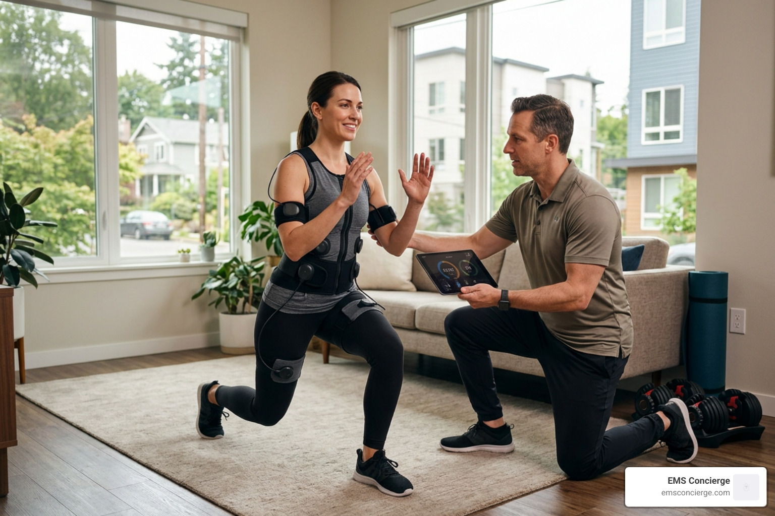
For healthy individuals, superimposing electrical stimulation onto voluntary exercises enhances neuromuscular recruitment. In an 8-week randomized trial of non-athletic adults, researchers evaluated the effects of core strengthening combined with wearable EMS garments (Yoo et al., 2023).
The group performing exercises with superimposed EMS showed significantly higher contracted muscle thickness across the rectus abdominis, external oblique, internal oblique, gluteus medius, and adductor longus compared to exercise alone ($p < 0.05$). However, whole-body fat percentage and functional performance tests (such as McGill's torso stability test and Functional Movement Screen scores) did not show statistically significant differences between groups.
This suggests that while EMS drives targeted neuromuscular adaptations, it works best when integrated into a structured, comprehensive physical regimen. Enhancing your weekly workout routine with supervised stimulation provides the precise electrode contact and progressive overload needed to translate these neuromuscular activations into practical fitness improvements.
Electrical Stimulation vs. Conventional Recovery Modalities
Muscle recovery encompasses many techniques, from active cool-downs to passive interventions. Understanding how electrical stimulation fits into this landscape ensures realistic expectations.
| Recovery Modality | Primary Mechanism | Impact on DOMS | Impact on Muscle Atrophy | Accessibility & Convenience |
|---|---|---|---|---|
| NMES / EMS | Motor nerve depolarization; muscle pumping | Low / Non-significant | High (preserves fiber size) | Moderate; requires specialized units or certified trainers |
| TENS | Sensory nerve stimulation; pain gating | Moderate (transient pain relief) | None (no contraction) | High; portable handheld consumer units |
| Active Recovery | Voluntary low-intensity movement (e.g., cycling) | Moderate (temporary relief via blood flow) | Low / Maintenance | High; requires time and access to equipment |
| Massage Therapy | Mechanical manipulation of soft tissue | Moderate (reduces perceived tightness) | None | Low to Moderate; requires a therapist |
| Compression | External hydrostatic pressure gradients | Low to Moderate (edema reduction) | None | High; wearable sleeves and boots |
Navigating these modalities helps determine when electrical stimulation serves as an effective tool. Exploring in-home EMS training options helps clarify how full-body stimulation platforms compare with standalone passive recovery gadgets.
Evidence-Based Protocols: Electrical Stimulation for Muscle Recovery After Exercise
Using electrical stimulation post-exercise aims to accelerate recovery, clear metabolites, and restore baseline muscle performance. However, clinical evidence shows that not all stimulation protocols deliver equal results.
High-intensity eccentric training causes structural micro-tears in sarcomeres, triggering inflammatory cascades and muscle stiffness. While low-frequency, non-contractile stimulation does not instantly reverse this structural damage, recent research exploring electromagnetic stimulation on autonomic recovery demonstrates that paired central and peripheral stimulation can improve autonomic nervous system balance (Keriven et al., 2025). Athletes receiving paired stimulation showed significant increases in heart rate variability (HRV) metrics—including high-frequency (HF) and low-frequency (LF) power—at 72 hours post-exercise, indicating faster parasympathetic reactivation.
Managing Delayed-Onset Muscle Soreness (DOMS) and Autonomic Recovery
Despite marketing claims that electrical devices "flush soreness away," scientific reviews urge caution. An extensive meta-analysis evaluating evidence on electrical stimulation for DOMS analyzed 14 randomized controlled trials ($n = 435$) and found that electrical stimulation was no more effective than sham devices or passive rest for resolving DOMS or restoring peak force capacity at 24, 48, 72, or 96 hours post-exercise.
Laboratory trials measuring systemic biomarkers—such as reactive oxygen species (ROS), interleukin-6 (IL-6), total antioxidant capacity, and blood lactate—showed no significant divergence between low-intensity non-contractile stimulation and sham controls.
In short, while electrostimulation does not instantly clear post-workout soreness, it remains an effective modality for autonomic balance, microvascular perfusion, and preserving neuromuscular pathways during recovery periods.
Optimizing Protocols, Safety, and Implementation
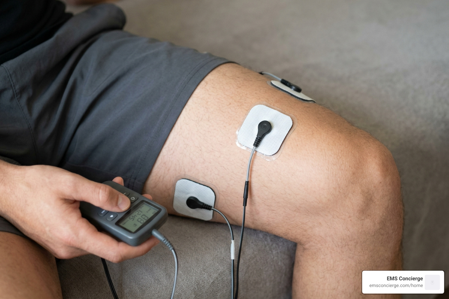
Applying electrical current safely and effectively requires adhering to established clinical parameters, precise electrode placement, and appropriate contraindication screening.
Stimulation Parameters: Frequency, Pulse Width, and Intensity
The physiological response to electrical stimulation depends on specific electrical parameters:
- Frequency ($text{Hz}$):
- 1–10 Hz (Twitch Mode): Elicits individual, non-fatiguing twitches. Ideal for low-threshold hemodynamic pumping and active recovery protocols.
- 20–50 Hz (Smooth Tetany): Produces a fused muscle contraction. Commonly used for endurance and early postoperative muscle re-education.
- 50–85 Hz (High-Frequency Tetany): Recruits high-threshold Type II fast-twitch motor units. Ideal for strength training, explosive power development, and limiting disuse atrophy.
- Pulse Duration (Pulse Width): Typically set between 200 and 400 microseconds ($mutext{s}$). Wider pulse durations engage deeper motor axons at lower overall voltage, improving comfort.
- Duty Cycle (On/Off Ratios): Continuous stimulation induces rapid neuromuscular fatigue. Effective protocols use intermittent duty cycles (such as 5–10 seconds of contraction followed by 10–50 seconds of rest) to allow metabolic recovery between contractions.
- Current Intensity: Should be adjusted to the maximum tolerable level without sharp pain. In clinical settings, higher training intensities correlate with greater preservation of muscle cross-sectional area.
Contraindications and Safety Best Practices
Before using any electrostimulation protocol, screen for key contraindications:
- Active Implanted Electrical Devices: Absolute contraindication for individuals with cardiac pacemakers, implantable cardioverter-defibrillators (ICDs), or deep brain stimulators.
- Pregnancy: Avoid placing electrodes over the abdomen, pelvis, or lower back.
- Epilepsy & Neurological Disorders: Electrodes must never be placed across the neck (carotid sinus region) or trans-thoracically across the heart.
- Compromised Skin Integrity: Do not apply adhesive pads over open wounds, active dermatological lesions, broken skin, or areas with acute sensory deficits.
- Active Malignancies & Deep Vein Thrombosis (DVT): Do not apply stimulation over regions with active tumors or suspected blood clots, as increased local blood flow may dislodge a thrombus.
Frequently Asked Questions About Electrical Muscle Stimulation
How does electrical stimulation promote tissue healing?
Electrostimulation encourages local tissue recovery primarily through hemodynamic and cellular pathways. By generating rhythmic twitches or microcurrent potentials, it increases capillary blood flow, enhances local oxygen extraction, and accelerates the clearance of metabolic waste. At the cellular level, specific frequencies stimulate ATP production within mitochondria and activate amino acid transport, supporting muscle protein synthesis and tissue remodeling.
Can electrical stimulation replace active recovery exercise?
No. Electrical stimulation serves as a valuable supplement to active recovery, not a replacement. While electrical stimulation induces muscle contraction without cardiovascular or articular strain, voluntary active recovery (such as light cycling or walking) stimulates full-body cardiovascular circulation, engages joint mechanoreceptors, and lubricates articular cartilage. Combining light voluntary movement with targeted stimulation delivers more balanced results than relying on passive modalities alone.
What are the main contraindications for electrostimulation?
The primary contraindications include implanted electronic devices (such as pacemakers or defibrillators), pregnancy (avoiding the abdominal and pelvic regions), active seizure disorders, acute deep vein thrombosis, open skin wounds, and placing electrodes across the anterior neck or thoracic cavity. Always consult a physician or physical therapist prior to beginning an electrical stimulation protocol if you have underlying medical conditions.
Conclusion
The clinical evidence for electrical muscle stimulation highlights its distinct capabilities. It is not an instant cure for post-exercise soreness, but it remains one of the most effective tools for preventing disuse atrophy, overcoming arthrogenic inhibition after joint surgery, and enhancing motor unit recruitment when paired with exercise.
By tailoring stimulation parameters—such as frequency, pulse duration, and intensity—to individual needs, electrostimulation bridges the gap between passive rest and active performance.
At EMS Concierge, we bring this advanced neuromuscular technology directly to you. Our certified trainers deliver in-home, personalized fitness sessions using professional-grade Miha Bodytec equipment, helping you achieve gym-level muscle recruitment in convenient 20-minute sessions. Whether you want to overcome muscle activation deficits or improve your weekly fitness routine, explore our specialized muscle recovery stimulation services to elevate your strength and recovery today.